


Otitis externa in dogs
Anita Patel BVM DVD FRCVS, RCVS specialist in veterinary dermatology explores the reasons why we don’t always succeed in preventing recurrence of otitis externa in dogs
Otitis externa in dogs is common and, in many, it can become a recurring problem. With time, the pathological changes to the ear canals will become irreversible; in these cases, the best alternative treatment is total ear canal ablation. To avoid this outcome, an understanding of the normal physiology and how to restore it by reversing the pathophysiological changes within the ear is paramount to successful long-term management.
Otitis is considered a multifactorial condition. The primary causes (see Table 1) must be managed in order to prevent recurrence; however, managing them alone will not necessarily prevent recurrences of the ear disease, nor its progression to a chronic state. Secondary infections, pathological changes to the ear canals and perpetuating factors also need to be addressed (see Table 1). These factors interfere with the normal physiological function and alter the anatomical structures within the ear making it more difficult to resolve. Progressive otitis externa can result in otitis media.
So, to understand how a normal ear functions and how anatomical changes result in ongoing disease, the following questions need to be addressed.
What is the self-cleaning mechanism?
This is the process by which the epithelial cells lining the ear canals and the tympanum turn over and migrate from the tympanic membrane towards the external auditory meatus. The process removes cerumen, cellular debris, microorganisms and allergens, thus maintaining a healthy microenvironment within the ear. Epithelial migration has been extensively studied in humans and rats, and epithelial migration, radially outwards from the tympanum, has been reported in dogs (Tabacca et al, 2011). Inflammation will disrupt this self-cleaning mechanism; the earlier it is restored the less likely it is to progress into chronic inflammatory disease with irreversible changes.
Why does this mechanism become disrupted?
Disruption usually stems from inflammation arising from primary causes, secondary infection, predisposing factors and perpetuating factors. The signs of inflammation: oedema, erythema, increased secretions and hyperplasia, lead to stenosis (see Figure 1), which interferes with the rate of cell turnover, drainage and subsequently the self-cleaning mechanism. Chronic, or recurrent, inflammation results in the progressive thickening of the epidermis and increased secretion and retention of cerumen within the ear canal lumen, all of which favour microbial growth (see Figure 2). This vicious cycle of increased thickening, changes in microbiome, further thickening, etc., leads to fibrosis and calcification of the cartilages making therapy more difficult.
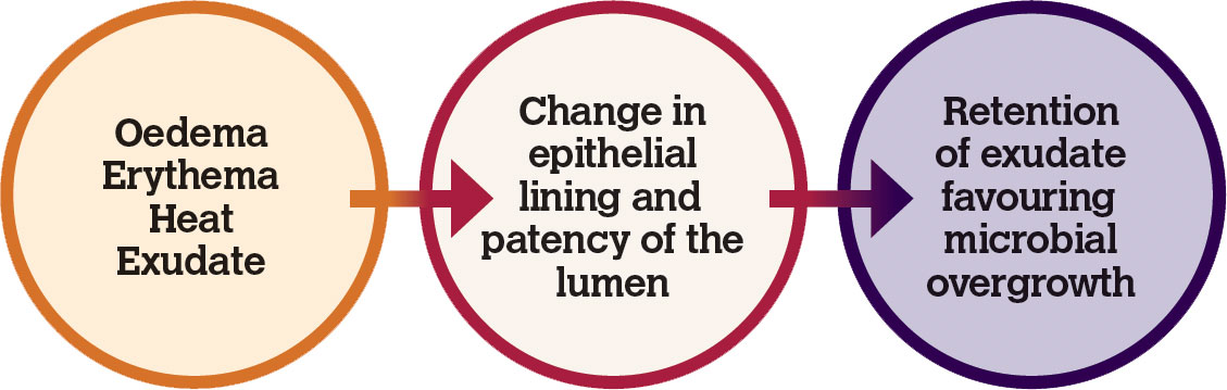
Figure 1: Pathophysiological changes lead to otitis externa.
How to restore the self-cleaning mechanism
Reversing the changes associated with inflammation, ie. oedema, erythema and exudation, and treating secondary microbial infections will restore the epithelial migration. Glucocorticoids are indicated to reverse the oedema, erythema and exudation. Antimicrobials are indicated for the infection. Once treated the epithelium should be smooth, glistening and pale (Figure 3). The sooner the swelling is reversed, and drainage re-established, the less likely it is to lead to chronic irreversible changes.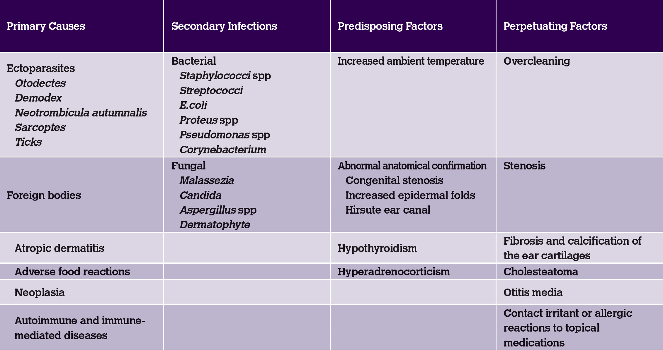
Table 1: Causes of otitis in the dog.
The preparations available in the veterinary market contain a combination of glucocorticoids, antibiotics and antifungals; however, they differ in their antibacterial and antifungal ingredients and in the level of potency of the glucocorticoid. The choice of medication depends on individual needs at the time. It may be necessary to use systemic glucocorticoids for a short period, for example five to 14 days, to reverse the inflammation rapidly, even though most of the ear preparations contain some form of steroid. On the practical side, systemic glucocorticoids help reduce swelling and subsequently the pain, making topical treatment easier for the owner to do.
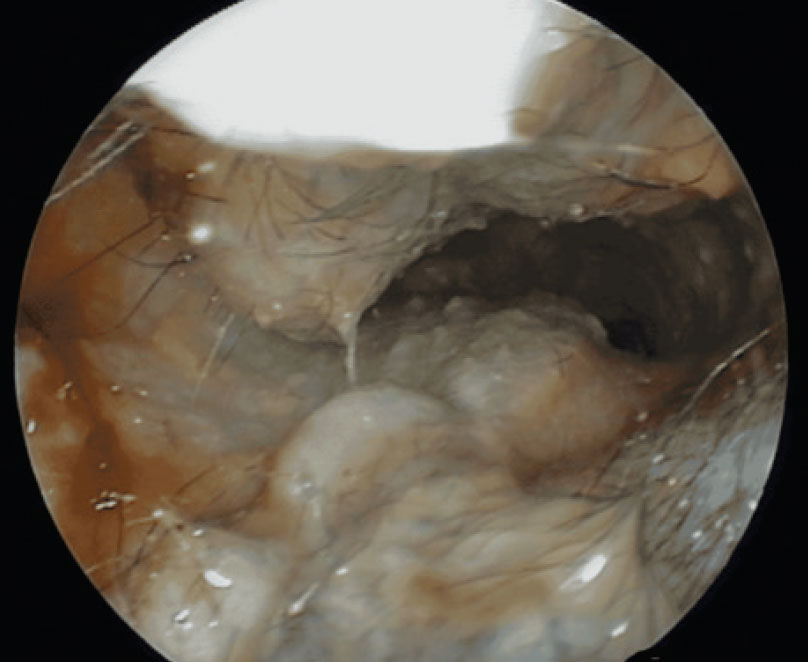
Figure 2: Inflammation, oedema, erythema, hyperplasia, stenosis and purulent exudate can be found in the vertical ear canal.

Figure 3: Post treatment, the epithelial lining should be smooth and pale.
Systemic antibiotics are not indicated as they have very limited value in resolving secondary infections in otitis externa. If further pain relief is needed it is probably better to add opioid analgesics combined with systemic glucocorticoids, as non-steroidal anti-inflammatory drugs have little effect on reversing the oedema.
Ear cleaning is an essential part of the therapeutic regime, whether done as home flushing alone, or following flushing in the clinic under general anaesthetic. Cleaning facilitates the removal of ceruminous debris, bacteria and bacterial toxins, pus and by-products of inflammation, which aids in restoring the microenvironment within the ear lumen. However, overcleaning can lead to maceration of the epithelium, preventing restoration of the self-cleaning mechanism and so perpetuating the otitis.
Summary
- Restoring the self-cleaning mechanism is essential to prevent recurrence of the otitis.
- Topical therapy is the mainstay of treatment of otitis externa.
- Glucocorticoids reduce inflammation and stenosis and often reduce discomfort.
- More severe or chronic cases may require systemic glucocorticoids.
- Systemic antibiotics have no, or very little, value in the management of otitis externa.
About the author
Anita Patel BVM DVD FRCVS, is a diplomate and a recognised RCVS specialist in veterinary dermatology. She has worked exclusively as a dermatologist for the last 15 years and lectures on all aspects of small animal dermatology in the UK, Europe, Africa and Asia.
- Barnard N and Foster A. 2017. Pseudomonas otitis in dogs: a general practitioner's guide to treatment. In Practice, 39, 386-398.
- Chester DK. 1998. Medical management of otitis externa. Veterinary Clinics of North America, 18, 799-812.
- Gotthelf LN. 2000. Small Animal Ear Diseases. W. B. Saunders Company, Philadelphia.
- Griffin CE. 1993. Otitis externa and otitis media. In: Current Veterinary Dermatology, eds CE Griffin, KW Kwochka and JM McDonald. Mosby Year Book, St Louis. pp. 245-262.
- Harvey RG, Harari J and Delauche AJ. 2001. Ear Diseases of the Dog and Cat. Manson Publishing Ltd, London.
- Nuttall T. 2016. Successful management of otitis externa. In Practice, 38, 17-21.
- Wilke JR. 1988. Otopharmacology. Veterinary Clinics of North America, 18, 783-812.