Focus - Companion animal - August 2019
Canine ophthalmic emergencies
Ocular emergencies can be daunting as they require rapid assessment and diagnosis. Treatment needs to be timely, appropriate and often aggressive to provide relief of pain and potentially save vision and the globe. In part one of this article, Natasha Mitchell MVB DVOphthal MRCVS, reviews globe proptosis, glaucoma, lens luxation and sudden onset blindness
Globe proptosis
Proptosis is forward displacement of the globe beyond the orbital rim, spasm of the eyelids prevents spontaneous return of the globe into the orbit (Figure 1a). Trauma of this nature is most often caused by road traffic accidents or dog attacks. It is much more common in brachycephalic breeds, as they have shallow orbits so proptosis may occur with relatively little trauma.
The eye needs to be kept moist from the beginning, even with tap water or Vaseline, before arrival at the clinic. Topical lubricant or antibiotic ointment should be applied as soon as the animal arrives at the vets. Once the patient has been treated for shock and head injuries, and stabilised, the extent of ocular damage can be assessed. Negative prognostic indicators include rupture of three or more extraocular muscles, globe rupture, extensive hyphaema, a negative dazzle reflex and a negative consensual pupillary light reflex (PLR). In this situation, enucleation may be the best course of action. However, globes that are intact and have some potential for salvage are best repositioned, and later they can be reassessed to determine if there is comfort and vision.
Prompt treatment is essential if the patient is stable, as the sooner the globe is replaced, the better the prognosis. General anaesthesia is required. A lateral canthotomy makes the procedure easier, but isn’t always necessary. Topical lubricating gel or antibiotic ointment is applied to the globe. The eyelid margins need to be pulled anteriorly over the equator of the globe, while some pressure is put on the globe at the same time to push it back into the orbit. Two methods can be used, depending on personal preference.
- The eyelids may be grasped and pulled outwards with Allis tissue forceps, attaching them perpendicular to the eyelid margin and at least 5mm in from the leading edge. Simultaneously, the globe is gently but firmly pushed back into the orbit with digital pressure or a scalpel handle. The lateral canthotomy is repaired and a temporary tarsorrhaphy is performed, as shown in Figure 1b. Two to three horizontal mattress sutures are placed through stents to reduce tension (Figure 2).
- Two to three horizontal mattress sutures could be pre-placed using stents, without tying knots. A scalpel handle can be placed across the cornea, gently but firmly pushing the globe back into the socket, while simultaneously pulling the preplaced sutures outwards, drawing the eyelid margins over the globe. The scalpel handle is withdrawn once the eyelids are in the correct position over the globe, and the sutures can then be tied.
Accurate placing of the sutures is important, to avoid suture material rubbing on the globe. Directing the needle through the ‘grey line’ or meibomian gland orifices is a useful landmark. The suture ends could be tied in long bows, so that they could be undone for future examination and replaced if needed. The temporary tarsorrhaphy protects the cornea from desiccation and the globe from repeat proptosis until the orbital soft tissue swelling subsides. Topical broad-spectrum antibiotics and atropine may be applied through a gap at the medial canthus. Systemic nonsteroidal anti-inflammatory drugs (NSAIDs) and antibiotics are also prescribed. An Elizabethan collar is required to prevent self-trauma.
The sutures are left in place for at least two weeks (and up to three weeks), at which time the globe can be reassessed (Figure 3). If the outcome is undesirable at this stage, enucleation may be indicated. Premature removal of sutures can result in corneal ulceration, as lagophthalmos may still be present and tear production may be suboptimal. Sutures could be retied if the palpebral reflex is still weak. On-going monitoring of tear production using the Schirmer tear test is recommended, and supplementation with artificial tears is usually required for months. Referral could be considered because a medial canthoplasty surgery is useful, particularly in brachycephalic breeds. For the post-proptosis eye, it reduces the size of the palpebral aperture, improving the blink, reducing corneal exposure and making repeat proptosis less likely. The unaffected eye could also have a medial canthoplasty surgery to reduce the size of the palpebral aperture and make the globe less vulnerable to proptosis.
Glaucoma
Glaucoma is a large, diverse group of painful and blinding disorders. They share the common feature that the IOP is too high to permit the optic nerve and retina to function normally. There are a range of clinical signs which include episcleral congestion, corneal oedema and mydriasis, along with vision deficits (Figures 4 and 5). IOP is sustained over 25mmHg for glaucoma to develop. Chronic glaucoma is not an emergency, and may present with an enlarged globe (buphthalmos), peripheral deep corneal vascularisation, corneal Haab’s striae (cracks in Descemet’s membrane), lens subluxation, retinal degeneration and optic disc cupping (Figure 6).
Primary glaucomas occur due to a structural or physiological abnormality within the aqueous outflow pathways. Secondary glaucomas are more common, and are caused by a different ocular or systemic disorder that impairs aqueous humor outflow. Examples include uveitis, lens luxation or neoplasia. Treatment depends on the type/cause and stage of glaucoma in order to provide appropriate and timely reduction in IOP with the goals to alleviate pain and to preserve as much vision as is possible. The higher the initial IOP and the longer it has been present, the less chance there is of preserving any vision. Sustained high IOP for 24-48 hours can result in permanent blindness. Glaucoma is a very serious condition and referral is recommended to establish the underlying cause and the best treatment regime, along with assessment of the risk in the fellow eye.
Emergency medical treatment includes:
- Prostaglandin analogues, such as 0.005% latanoprost (Xalatan, Pfizer) and 0.004% travoprost (Travatan, Novartis Pharmaceuticals UK Ltd). Topical treatment increases uveoscleral outflow. They result in potent miosis and they are pro-inflammatory, and so exacerbate uveitis. They are contraindicated with anterior lens luxation and uveitis. One drop is applied twice daily.
- Osmotic agents. Mannitol 20% can be administered intravenously at a dose rate of 1-2g/kg over 20-30 minutes and rapidly causes reduction in IOP through dehydration of the vitreous and aqueous (and the patient). It can be repeated after six hours if necessary. It shouldn’t be used in cases of congestive heart failure, pulmonary oedema or any renal compromise, and therefore urine and blood tests for renal function are recommended prior to use where possible. It is a temporary measure to reduce the IOP, typically used while arranging for a referral if the prostaglandin analogue has not decreased the IOP within 30 minutes.
- Carbonic anhydrase inhibitors, such as 20mg/ml dorzolamide (Trusopt, MSD) and 10mg/ml brinzolamide (Azopt, Alcon). Topical treatment reduces aqueous production. They can be used even when there is lens luxation and uveitis. One drop is applied three times daily.
- Pain relief. Systemic opioids and NSAIDs are used until the IOP is controlled. However, systemic dexamethasone could be used as an alternative to NSAIDs if the eye appears very inflamed, if appropriate to the individual case.
- Aqueocentesis may be required if these emergency methods fail, usually performed by an ophthalmologist.
Anterior lens luxation
Anterior lens luxation is the displacement of the lens from its normal position into the anterior chamber. Dislocation of the lens may be due to a primary inherited defect in the ciliary zonules in certain breeds, including many terrier breeds, or secondary to chronic glaucoma, blunt trauma, degenerative changes in older dogs or intraocular neoplasia. Typically, it causes a rapid elevation of IOP, which is potentially blinding.
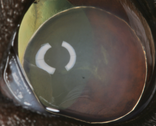
The lens may be visualized within the anterior chamber if it is anteriorly luxated, and the eye will be sore (Figure 7). The lens is clear with an acute luxation, and gradually develops cataract (Figure 8). Corneal oedema develops particularly where the lens is resting against the endothelium, usually centrally and ventrally (Figure 9). The other eye should be examined as it is a bilateral condition when primary. The IOP should be measured and potential vision assessed with the dazzle reflex and consensual PLR. If there is doubt as to the position of the lens, for example if corneal oedema is pronounced, transcorneal ultrasound offers a safe and reliable inspection of the globe and the lens can be easily imaged. Primary acute anterior lens luxation is a true emergency and is ideally referred to an ophthalmologist urgently, where surgical removal of the lens, along with treatment of the fellow eye, can be discussed. Pain relief is required. Topical 1% tropicamide or 1% atropine can be used to attempt to relieve the pupil block and therefore reduce the IOP. However, both mydriatics cause significant increases in IOP with angle closure glaucoma, so caution is advised. Topical prostaglandin analogues need to be avoided as they would cause pupil block due to miosis, but topical carbonic anhydrase inhibitors are appropriate. Trans-corneal reduction (couching) of anterior lens luxation in dogs with lens instability has been reported (Montgomery et al, 2014). After this procedure, topical prostaglandin analogues are used twice daily to keep the lens in the vitreous chamber and to reduce the IOP. With secondary anterior lens luxation, if there is no potential for vision, enucleation may be the best option for the patient.
Sudden onset blindness
Dogs with sudden onset vision loss may have a visible opacity in the ocular media, such as hyphaema, diabetic cataracts or signs of uveitis, glaucoma or retinal detachment. A history and ocular examination should lead to a diagnosis.
It is much more challenging to be presented with a dog with amaurosis, which is blindness without any apparent ocular lesion. The main differential diagnoses are:
- Sudden acquired retinal degeneration syndrome (SARDS);
- Retrobulbar optic neuritis;
- Neoplasia of, or adjacent to, the optic chiasm; and
- Central blindness.
SARDS can be differentiated from the other neurological conditions definitively by an electroretinogram. There is a higher index of suspicion in a middle-aged to older dog presenting with some combination of polyuria, polydipsia, polyphagia, enlarged abdomen and panting, with a fundus that appears normal. The pupils are dilated, and initially they are slowly responsive to bright white light, unresponsive to red light, and more strongly responsive to blue light stimulation. The aetiopathogenesis remains unclear, and it may be an endocrine or neuroendocrine disorder, a stress response, an autoimmune retinopathy, or there may be more than one aetiology. There is no successful treatment for return of vision.
Optic neuritis can be caused by infectious disease, granulomatous meningoencephalitis (GME), optic nerve neoplasia or it can be idiopathic. PLRs are absent and there may be some visible changes to the fundus including optic disc oedema (blurring), loss of the physiological cup and haemorrhages on the disc or within the adjacent retina. Lymphoma, pituitary carcinoma, paranasal sinus carcinoma and meningioma have been associated with sudden onset blindness with reduced or absent PLRs but normal fundus appearance. PLR is subcortical, so it is not affected in the case of central blindness. This can arise after general anaesthesia due to cerebral hypoxia, post seizure, after severe head trauma or due to GME.
It can be difficult to reach a diagnosis with clinical examination alone, and in many cases of amaurosis, further testing with an electroretinogram, magnetic resonance imaging and cerebrospinal fluid analysis may be necessary before a diagnosis can be made.
Part two, which will feature in the next issue, will cover trauma including eyelid laceration and corneal laceration, foreign bodies, melting ulcers and descemetocoeles.
BSAVA Manual of Canine and Feline Ophthalmology. 2014. Third Edition. Edited by David Gould and Gillian McLellan. ISBN 978-1-905319-42-8
Handbook of Veterinary Ophthalmic Emergencies. 2002. First Edition. David Williams & Kathy Barrie. Edited by Thomas Evans. ISBN: 978-0-7506-3560-8
1. After surgical replacement of a proptosed globe, a temporary tarsorrhaphy should be left in place for:
a. 24 hours
b. 1 week
c. 2-3 weeks
d. At least a month
2. Which of the following patients with proptosis would be expected to have the best prognosis for salvage of globe and vision?
a. A young domestic shorthair after RTA in the past hour
b. A young Shih tzu with medial strabismus and lagophthalmos
c. A young Pug with hyphaema and a negative dazzle reflex
d. A young Sheltie with zygomatic arch fracture
3. Glaucoma occurs after sustained intraocular pressure above what value?
a. 10mmHg
b. 15mmHg
c. 25mmHg
d. 50mmHg
4. Which of the following clinical features are present in acute glaucoma?
a. Episcleral congestion
b. Buphthalmos
c. Haab’s striae
d. Lens subluxation
5. Clinical signs of anterior lens luxation include:
a. Sudden onset blindness and amaurosis
b. Normal menace response but negative dazzle reflex
c. Gradual onset blindness with very painful eye
d. Corneal oedema ventrally and centrally


